Basal cell carcinoma
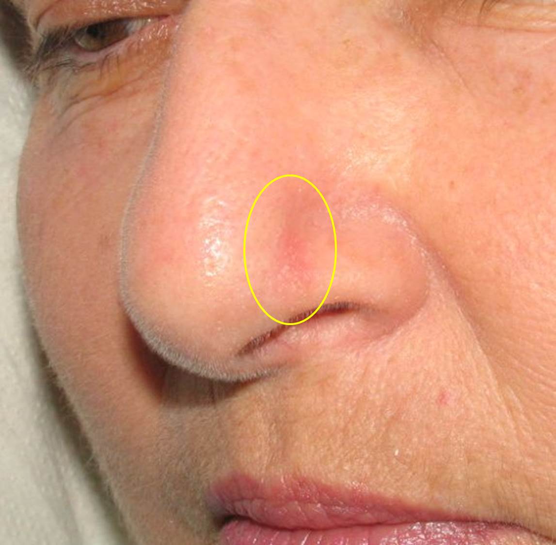
Basal Cell Carcinoma | BCC – Basal Cell Carcinoma
BCC is the most common skin cancer in the world. It is generally believed that in most cases the cause is ultraviolet (UV) radiation from sunlight, although additional factors certainly exist. A well‑known and common cause in Israel is the appearance of BCC tumors in patients who were treated many years ago with radiation for skin conditions such as tinea capitis (scalp fungal infection), acne, and others. Today, the mutation in the gene responsible for the malignant behavior of the tumor is known; it was identified in patients with a congenital mutation causing Gorlin syndrome, who develop multiple BCC tumors. The incidence of the disease is steadily rising, likely due to increased time spent outdoors and damage to the ozone layer, which allows greater penetration of UV radiation through the atmosphere.
BCC usually does not metastasize or spread through the bloodstream or lymphatic system to other organs. Instead, it spreads by infiltrating and destroying adjacent tissues, and the larger it becomes, the harder it is to remove. Therefore, early treatment is important.
How does the tumor appear? BCC typically appears on sun‑exposed areas such as the face, scalp, ears, neck, chest, back, and legs. It can present in several forms. The most common is a dome‑shaped, pearly or translucent bump, often with visible dilated blood vessels. Another common form is a wound that repeatedly recurs in the same spot, seemingly healing each time but never fully resolving. Superficial BCC, an early form of the tumor, appears as a shiny pink patch on the skin. A less common but more aggressive form, called morpheaform, appears as a waxy, white‑yellow scar‑like lesion. Some BCC tumors that develop after radiation have a distinct pigmented appearance.
What damage does the tumor cause? Except for extremely rare exceptions, the tumor does not metastasize, but it does not disappear on its own. It continues to grow locally, causing bleeding and tissue destruction unless removed. When located near vital structures such as the eyes, ears, nose, or near a nerve, the potential damage can be severe unless treated early.
Biopsy Any skin lesion or wound that does not heal must be evaluated by a dermatologist. If the clinical diagnosis is unclear, a biopsy is recommended. A biopsy is a simple procedure, usually performed in the clinic under local anesthesia, in which a small tissue sample is taken for microscopic examination. In some cases, the doctor may choose to remove the entire lesion immediately without waiting for biopsy results.
Treatment Once diagnosed as BCC, several treatment options exist:
- Standard surgical excision – A small surgery in which the lesion is removed completely with a margin of approximately 0.5 cm of clinically normal skin. The wound edges are sutured using dermatologic surgical techniques. The removed tissue is examined by a pathologist to determine the tumor type and whether it was fully excised.
- Electrodessication and curettage – The tumor is scraped with a curette (a sharp spoon‑like instrument), and the base is treated with electric cautery.
- Cryosurgery – Deep and thorough freezing of the tumor and surrounding tissue using direct liquid nitrogen spray.
- Topical therapy – Application of medications such as Efudix (5‑fluorouracil) or Aldara (imiquimod), which activates the patient’s immune system to treat the tumor. This is suitable mainly for superficial tumors.
- Mohs surgery – A specialized surgical technique performed by a dermatologic surgeon trained in Mohs. The tumor is removed under local anesthesia, and the tissue is examined under a microscope by the surgeon to ensure complete removal. Additional layers are removed as needed until all cancerous tissue is cleared. This method yields very high cure rates and is recommended for recurrent tumors, partially removed tumors, large tumors, or tumors in critical areas such as the eyes, ears, nose, and mouth. After complete removal, the wound is closed using aesthetic reconstruction techniques or left for gradual secondary healing.
- Radiotherapy – Electron beam radiation tailored to target tumor cells in the skin. Experts consider it highly effective, though controlled studies are limited. Radiation dose is adjusted to tumor location, surrounding tissues, and patient tolerance. Treatment is divided into daily fractions to reduce damage to healthy tissue. A lead shield is used to protect unaffected skin. Radiotherapy is especially suitable for patients at high surgical risk, patients with multiple tumors, those fearful of surgery, or tumors in locations where surgery may cause significant damage. However, the need for multiple hospital visits can be a limitation. Side effects include acute skin burns, wounds at the tumor site, dryness of nearby mucous membranes, and late effects such as sensitive skin, slow healing, and a small risk of radiation‑induced secondary cancers many years later. Radiotherapy is included in Israel’s national health basket.
- Photodynamic therapy (PDT) – A relatively new treatment in the national health basket. It is intended for sun‑damaged areas and superficial BCC. A photosensitizing agent is applied and absorbed preferentially by abnormal cells. After about 30 minutes, the area is exposed to red‑spectrum visible light, converting the agent into reactive oxygen species that destroy the affected cells. The skin reacts like a sunburn, and within days the damaged cells peel off, leaving the skin clearer. Some claim that incomplete treatment may make tumors more aggressive, but this has not been proven.
- Biologic therapy – These treatments were developed after identifying the mutation responsible for BCC. Unlike systemic chemotherapy (not used for BCC), these oral medications target the biological pathway driving tumor growth. The drug Erivedge is included in Israel’s health basket (another drug is expected soon). It is very expensive and reserved for cases where surgery or radiation is not possible.
Will there be a scar? Since most tumors appear on facial skin, many patients understandably worry about cosmetic results. Small tumors treated conservatively often yield excellent cosmetic outcomes. In cases requiring Mohs surgery, various aesthetic reconstruction options exist, usually producing excellent results. Even when the immediate postoperative appearance is not ideal, local treatments can often improve the outcome.
Follow‑up Studies show that once a person develops BCC, there is a 40% chance of developing another within 5 years. Therefore, regular dermatologic follow‑up is essential. Patients must be alert to any non‑healing wound. Consistent use of sunscreen and sun protection is crucial.
Associated risks Patients with multiple BCCs or other skin cancers such as SCC have an increased risk of melanoma. BCC itself cannot transform into melanoma.
Prevention Because BCC is often caused by UV radiation, sun avoidance and protection can help prevent it. Protection is especially important in children. Patients should perform frequent self‑exams and undergo a full‑body skin check at least once a year.
Seek shade during peak sun hours (10:00–16:00). Wear light‑colored, tightly woven clothing, long sleeves, long pants, a wide‑brimmed hat, and sunglasses. Use broad‑spectrum sunscreen (UVA + UVB) with SPF 15 or higher, and reapply every two hours — even on cloudy days.
Patients at high risk should see a dermatologist two to three times a year, preferably the same doctor who knows their skin and has experience diagnosing these tumors.
Dr. Gilead provides structured follow‑up for high‑risk patients using dermoscopy. Preventive treatments are performed when needed, and new tumors are removed using Mohs surgery, standard excision, or referred for other appropriate treatments.